Your browser is stacked with gallery tabs, and a handwritten list of structures sits beside the keyboard, half crossed out. Every photograph shows creases under museum lighting, and not one caption says whether the specimen was fresh or fixed. You still cannot picture the seahorse-shaped part tucked into the inner side of each temporal lobe. What would it take to hold that curve in your head once and stop starting over?
You are several pages into the image results and still unsure what you are looking at. Some photographs show a dark brown specimen under museum lighting, others show pale gray shapes with no surface at all, and the captions skip the detail that would tell you which is which. Here is the thing itself, plainly. A real human brain is soft and wet, grayish where the folds crowd across the surface, paler inside where it has been cut. Press a thumb into it and the print stays a moment. The folds are the point: they pack a wide sheet of tissue into a space that has to close inside a skull, which is why most of what a photograph shows is crease rather than flat. One caution before the anatomy. Published work on what a real brain looks like is thin, and a useful study would have to measure the same things the same way: weight and volume before and after preservation, fold depth, and the size of the seahorse-shaped part in the same people over time.
The part shaped like a seahorse
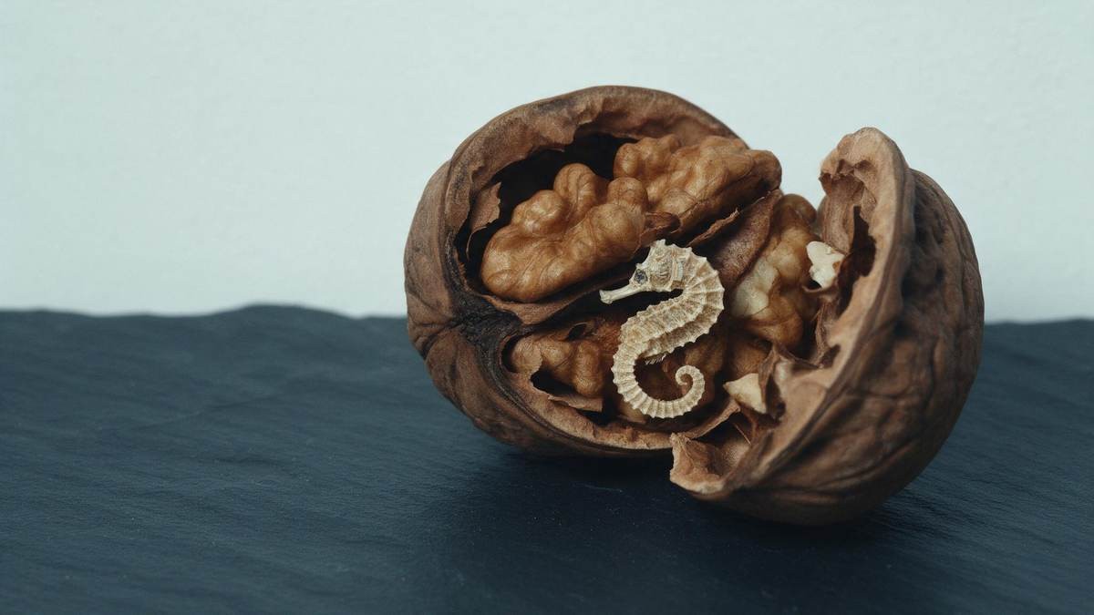
The hippocampus is small, curved along one axis like the seahorse it was named for, and tucked into the inner side of each temporal lobe. It sits so deep that no photograph taken from outside reaches it. New experiences are laid down there first, before they are stored elsewhere, which is why this structure is needed to form new memories and why it is studied so closely as memory changes with age.
How the tissue looks in each state
| State | What you see |
|---|---|
| Living brain | Pinkish-gray, blood-wet, soft enough that handling leaves marks |
| Freshly removed | Grayer, the edges beginning to relax under their own weight |
| Preserved brain | Firmer, duller, holding the shape it was fixed in |
| Brain in a scan | No color at all, gray levels standing for tissue, taken through the intact skull |
One on each side, mirrored like commas curled inward. Neither appears in a picture of the whole organ, which is why you keep meeting it in drawings instead. Ask for it by name in an image gallery and the results turn to diagrams.
The cost lands the next time someone asks you to describe it. You have worked through a run of galleries and still cannot say whether the specimen in front of you was fresh or fixed, because no caption mentioned it. So you go back to the first page of results and start again, comparing folds that were never meant to be compared, and you close the tab without looking at any single photograph long enough to hold the hippocampus in your head. The search repeats, the description never arrives.
 New memories and the hippocampus?Why can’t a photo reach itWatch the video while the curve is still on screen and the specimen stays in front of you
Why the folds matter more than the weight
People quote a figure for weight and move on, as though it settled what a brain is. What the figure hides is the surface. Brains of similar weight can differ in how their folds run, how deep the grooves sit, and how much of the sheet is tucked out of sight.
The pattern is not a template. Fold depth and spacing vary from person to person, so a photograph of one brain documents that brain. Diagrams smooth the variation out, which makes them useful for names and misleading for texture.
The choice depends on the question in front of you. You want the name of a structure, use the drawing. You want the sheen of the tissue, the grain, the way light pools in a groove, use the photograph, and check first whether the specimen was fresh or fixed.
The curve of the hippocampus is hard to picture from stills, so we walked around a specimen with a camera and you can watch the full video while the structure is still in front of you.
Living brain and preserved brain
Most pictures you will find are of preserved specimens, which is why people expect the real thing to be dark, firm and dry. It is not. Preservation changes color and firmness so a specimen can be handled, photographed and cut without collapsing.
Living brain: pinkish-gray, blood-wet, soft enough that handling leaves marks. Freshly removed: grayer, the edges beginning to relax under their own weight. Preserved brain: firmer, duller, holding the shape it was fixed in. Brain in a scan: no color at all, gray levels standing for tissue, taken through the intact skull.
Read the caption before the picture. A gallery that never says whether a specimen was fixed or fresh describes neither, and you end up judging a fixed specimen against a scan and drawing a conclusion about the organ.
What a photograph cannot tell you
A still frame carries shape and texture. It carries no weight, no firmness, no method. Without the protocol behind it, an image cannot tell you how the tissue was handled, how long it sat in fixative, or why it was chosen for the gallery.
So the careful account stops at description. Until measurements of the same brains, taken the same way before and after preservation, are published in quantity, there is nothing solid to put beneath the folds and the weight.
Galleries fill the gap with confident figures and no method attached. Treat an unlabeled number as decoration, then ask what was weighed, when, and in what state the tissue was in. The answer changes with that state, which is the whole point.
Underneath the anatomy is the question this page leaves open: what an adult can do in ordinary days so that a name, a recipe or an appointment arrives when it is wanted. The research on that is uneven, and it does not yet sort a useful habit from a fashionable one.
Does it change with age?
Everything changes with age, the brain included. People notice they take longer to learn something new or to pull a name back, and misplace things now and then. Forgetting where you put your keys or whether you locked the front door happens at any age, and on its own it is mild forgetfulness.
The line that matters is function. Serious memory problems make everyday things hard: driving, using the phone, finding your way home. Asking the same questions over and over, getting lost in places you used to know well, trouble following recipes or directions, confusion about time, people or place. Those are reasons to talk with a doctor.
Memory trouble also has more than one cause: depression, sleep problems, side effects of medicines, thyroid, kidney or liver problems, head injury, alcohol, low levels of vitamin B12. Finding the cause comes before any plan. If changes persist after a few weeks or keep worsening, see a doctor.
A photograph stops at the surface, and the film goes past it, taking the same question with it: watch the full video.
Everything on Everyday slips →
▶ Watch the memory video